What does a normal tympanic membrane look like?
The normal tympanic membrane is in the neutral position (neither retracted nor bulging), pearly gray, translucent and responding briskly to positive and negative pressure, indicating an air-filled space.
What does a healthy eardrum look like through an otoscope?
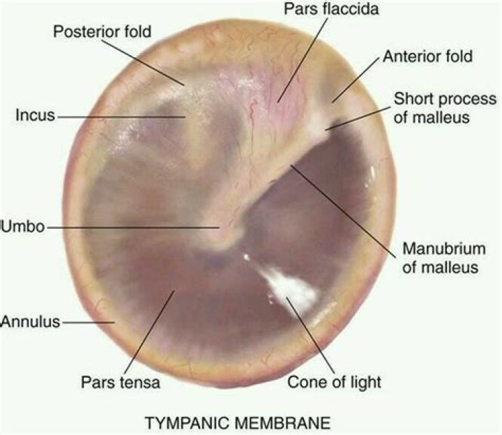
A healthy eardrum is concave inwards. The cone of light is seen on a healthy eardrum – it is from otoscope light reflected from the concave surface. The cone of light extends from the middle of the eardrum to the periphery.
How do you identify the tympanic membrane?
1) Color/shape-pearly grey, shiny, translucent, with no bulging or retraction. 2) Consistency – smooth. 3) Landmarks.
What does fluid behind eardrum feel like?
In general, symptoms of fluid in the ears may include: Ear pain. Feeling like the ears are “plugged up” Increasing ear pain when changing altitude, and being unable to “pop” the ears.
What are the parts of the tympanic membrane?
The tympanic membrane is comprised of three layers of tissue: the outer cutaneous layer, the fibrous middle layer, and a layer of mucous membrane on its innermost surface. The membrane is held in place by a thick ring of cartilage, a tough but flexible kind of tissue.
Where is the tympanic membrane located in the ear?
The tympanic membrane is a very thin structure that separates the outer ear canal from the middle ear space.
What color is tympanic membrane?
..:: The Tympanic Membrane ::.. 1) Color/shape-pearly grey, shiny, translucent, with no bulging or retraction. 2) Consistency – smooth.
How should an eardrum look?
Normal: The eardrum is pearly white or light gray, and you can see through it. You can see the tiny bones of the middle ear pushing on the eardrum. You see a cone of light, known as the “light reflex,” reflecting off the surface of the eardrum.
What is the normal appearance of the tympanic membrane?
The diameter of the TM is about 8-10 millimeters. Its outer surface is slightly concave. Tympanic membrane (TM) as continuation of the upper wall of external auditory canal (EAC) with angle of incline up to 45 degrees on the border between middle ear and the EAC.
What instrument is used to examine the tympanic membrane?
A modern electric otoscope/auriscope with its own light source is primarily used to examine the ear. An otoscope also has its own magnification, which gives a good view of the tympanic membrane (TM). Batteries need to be fully operational to allow optimal light during examination.
What is the tympanic membrane commonly known as?
The tympanic membrane is a vital component of the human ear, and is more commonly known as the eardrum.
How many layers comprise the tympanic membrane?
The tympanic membrane is comprised of three layers of tissue: the outer cutaneous layer, the fibrous middle layer, and a layer of mucous membrane on its innermost surface. The membrane is held in place by a thick ring of cartilage, a tough but flexible kind of tissue.